

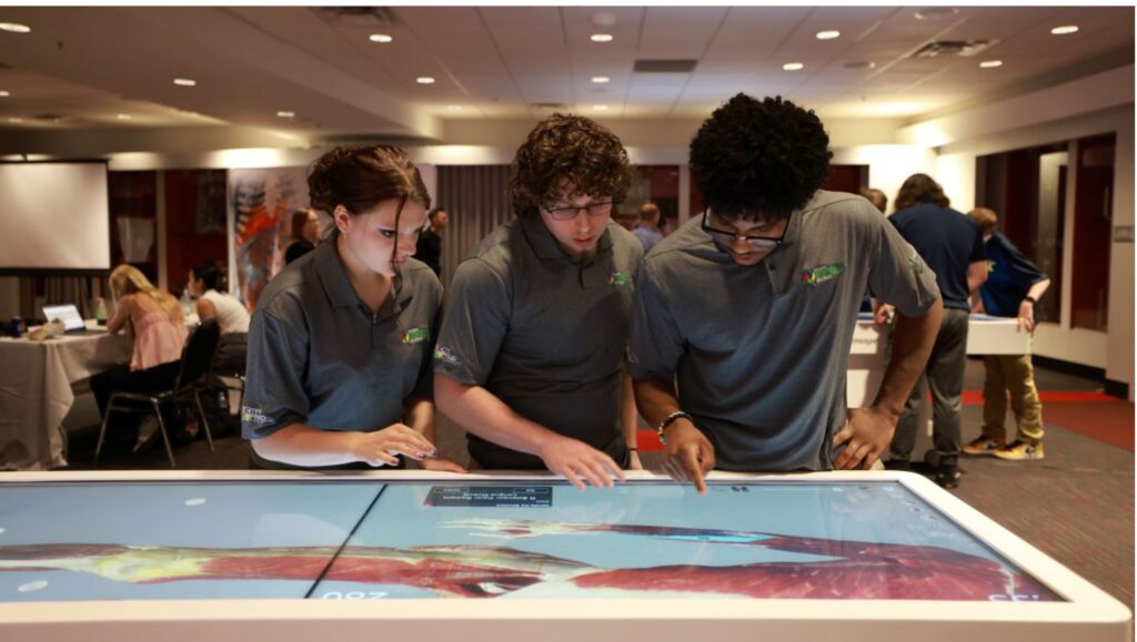
Our school is set to revolutionize the way students learn anatomy and other sciences with the arrival of the Anatomage Table. This state-of-the-art, interactive dissection tool allows students to explore and study the human body in 3D, providing a hands-on learning experience. The Anatomage Table uses advanced imaging technology to simulate life-sized, virtual cadaver dissections, complete with detailed layers of muscles, bones, and organs. With just a swipe of the hand, users can explore different bodily systems, zoom in on specific structures, and rotate images for a comprehensive view. Not only does the Anatomage provide a more in-depth look into human anatomy, it can also simulate chem labs, simulate a stroke, and even provide detailed anatomy of animals as well.
In an interview with Principal Mr. Channer, he confirmed that the Anatomage Table will be available to all students, including those in the art department, who can benefit from using it to understand anatomy better for their projects. “We want this incredible tool to be accessible to everyone,” said Mr. Channer, emphasizing that the table’s availability would not be limited to just science courses.
To prepare for the table’s rollout, teachers are set to begin training sessions in the coming weeks, ensuring that the table will be fully integrated into the curriculum by next semester. Some faculty members are even taking specialized training to enhance their teaching capabilities. For example, Mr. Bayer is undergoing additional training in chemistry to utilize the Anatomage Table’s features that intersect with chemical processes in the human body.
The introduction of the Anatomage Table marks a significant step in providing cutting-edge educational tools that cater to a wide range of learning experiences, making complex subjects more engaging and accessible to students across different classes and even schools.